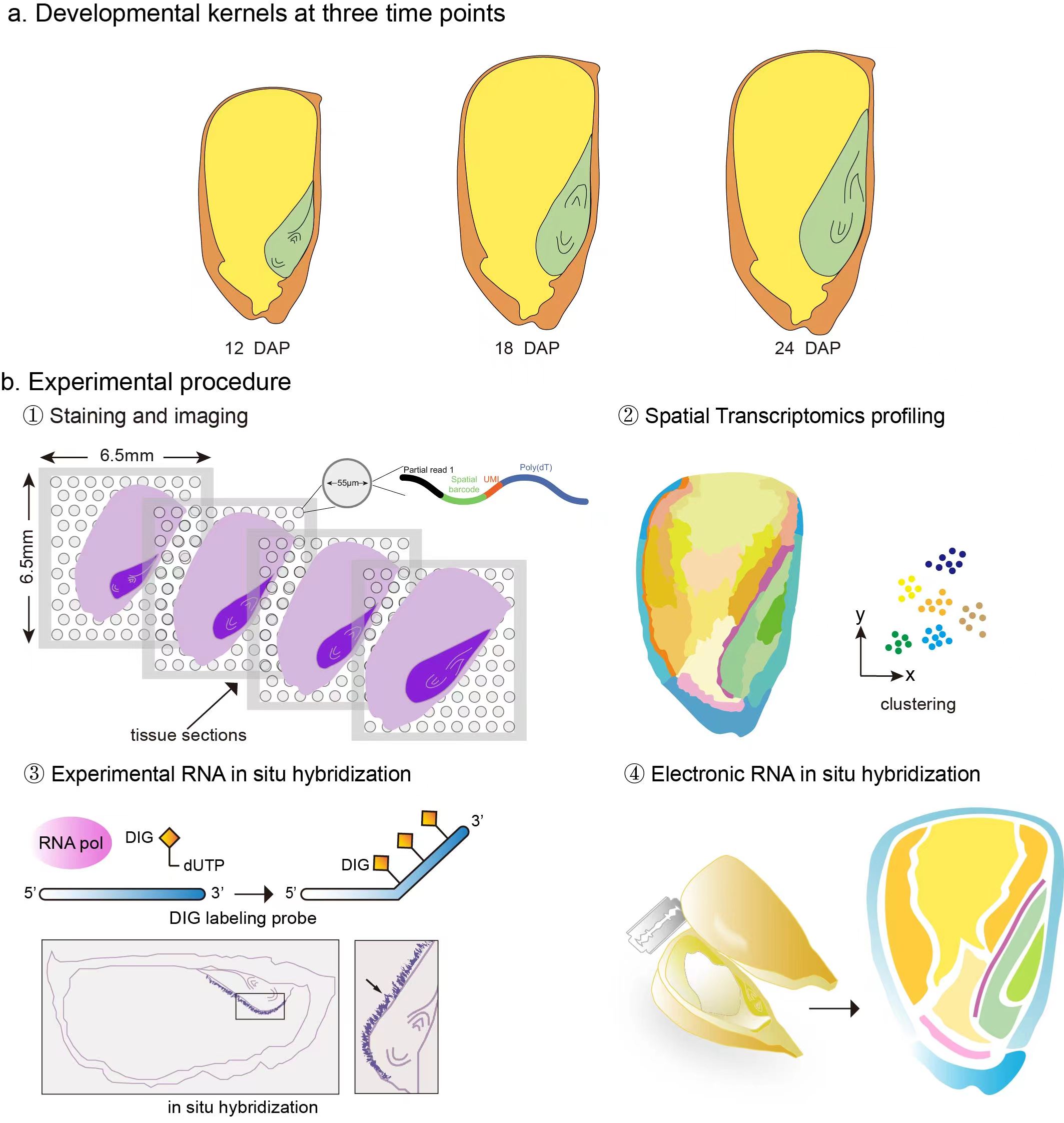
This resource accompanies a spatial transcriptomic analysis of maize kernels across grain filling, providing electronic RNA in situ visualisation for genome-wide gene expression.
How to cite
Fu, Y., Xiao, W., Tian, L. et al. Spatial transcriptomics uncover sucrose post-phloem transport during maize kernel development. Nature Communications14, 7191 (2023). https://doi.org/10.1038/s41467-023-43006-7
Please cite the original article when using this atlas or its visualisations. The article is open access under CC BY 4.0.
Biological question
How is sucrose distributed after phloem unloading?
The maize kernel brings together maternal tissue, filial endosperm and embryo. During filling, the transfer of carbon through the maternal–filial interface supports the accumulation of starch and protein in endosperm and lipids in embryo.
By combining microscopic sectioning with spatial transcriptomics, the study identifies the spatial organisation of kernel cell populations and gene programs that support nutrient delivery and reserve formation.
Spatial transcriptomic workflow used in the study
Atlas outputs
Electronic RNA in situ maps
Gene expression is visualised in tissue context, enabling exploration of marker genes and spatial patterns without requiring experimental in situ hybridisation for every target.
Key finding
Post-phloem transport
Maternal placento-chalazal tissue and the basal endosperm transfer layer form specialised interfaces that support nutrient uptake and downstream storage programmes.